<img alt="" src="https://secure.soil5hear.com/223550.png" style="display:none;">
Skip to content

Preclinical Histology & IHC Services

Industry-leading CAP/CLIA-certified preclinical and clinical histology services

Tumor Microarray H&E staining slides.

Custom Developed Histology & IHC Services

Champions leads the industry in pathology expertise and innovative automated technology providing you with the highest quality results.  As an endpoint for both preclinical and clinical studies, Champions' dedicated laboratories are fully equipped to support the entire sample lifecycle, including collection, processing, storage, staining for histology analysis, imaging, and specialized scoring by a board-certified pathologist; allowing the ability to process and store samples for histology, and IHC, as well as create custom tumor microarrays (TMAs).  

  • Expertise in preclinical sample analysis, including complex sample analysis needed for PDX models
  • GCLP-compliant clinical sample preparation and analysis
  • Over 200 custom developed protocols and fully optimized antibodies for research

Confidently Select TumorGraft® Models through Specialized IHC

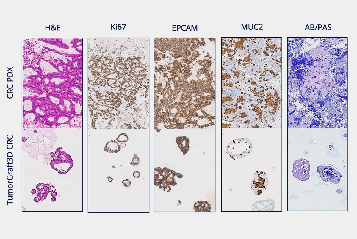
Champions provides standard and custom grossing, FFPE generation, H&E staining, marker optimization for IHC, scoring by board-certified pathologists, slide digitization and automation, and TMA creation and analysis (organized by indication and collated from over 800 well-characterized TumorGraft models) for target expression evaluation.  

View Preclinical Optimized IHC Antibodies

IHC results of PDX and TumorGraft3D models for research provided by IHC services.
Clinical Specialty testing services of IHC for clinical trial using PD-L1 CLIA test.

Evaluate Target Expression in Clinical Patient Samples With Our Histopathology Services

Champions Histopathology Laboratory has been CAP-accredited and maintains GCLP compliance. These added levels of compliance lead to additional scientific rigor and give you high-quality results to drive clinical trial success. 

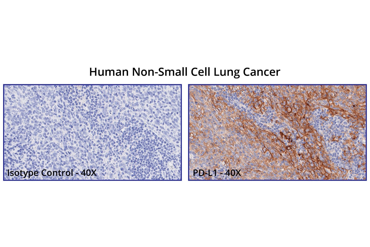
In addition, we have received CLIA certification, 21D2030870, which gives our laboratory the ability to perform PD-L1 Immunohistochemistry (IHC) testing on human tissues and cells from clinical trials.

Champions' Clinically Evaluated IHC Antibodies


Champions' histology and immunohistochemistry services are custom-developed and fully optimized to meet your needs in preclinical research. Champions leads the industry in pathology expertise and innovative automated technology that provides you with the highest quality endpoints complimentary to in vivo and ex vivo study results. 


Tab Imges_Medium31


Ready to start working with Champions Oncology? 
Frequently Asked Questions
What are preclinical histology services?

Preclinical histology services involve the preparation, examination, and analysis of tissue samples from animal models in the early stages of drug development. These services provide detailed insights into the effects of potential therapeutic compounds on tissue structure and function. By utilizing techniques such as tissue sectioning, staining, and microscopic analysis, preclinical histology helps researchers examine target expression at the level of the tissue of interest, which can aid in appropriate model selection for ex vivo and in vivo studies, and helps understanding the safety and efficacy of new drugs.

What is the role of histopathology in preclinical research?

Histopathology plays a critical role in preclinical research by providing detailed information about the microscopic structure and pathology of tissues in response to experimental treatments. It helps identify cellular changes, such as inflammation, necrosis, or tumor formation, that occur due to drug exposure. Histopathology is essential for evaluating target expression and assessing the potential toxicity of new compounds and understanding their mechanism of action.

What are the advantages of using preclinical histology services?

Using preclinical histology services offers several advantages, including:

  • Expertise: Access to specialized knowledge and skills in tumor tissue preparation and analysis.
  • Advanced Technology: Utilization of state-of-the-art imaging and optimized staining techniques for precise results.
  • Efficiency: Outsourcing histology tasks can accelerate project timelines by leveraging dedicated resources.
  • Quality Control & Assurance: Ensures consistent and high-quality results that meet regulatory standards, supporting the credibility of preclinical findings.
What Types of Imaging Technologies Are Used in Histology?

Histology utilizes various imaging technologies to visualize and analyze tissue samples, including:

  • Light Microscopy: Commonly used for examining stained tissue sections at high resolution.
  • Fluorescence Microscopy: Allows for the visualization of specific molecules within tissues using fluorescent tags.
  • Confocal Microscopy: Provides detailed 3D images of tissues by scanning multiple planes within a sample.
  • Electron Microscopy: Offers ultra-high resolution images of tissue ultrastructure, revealing cellular details at the nanometer scale.
How Do You Handle and Store Tissue Samples for Histology?

Handling and storing tissue samples for histology involves careful procedures to preserve tissue integrity and prevent degradation. Samples are typically fixed in a preservative like formalin shortly after collection to stabilize the tissue structure and then embedded in paraffin wax sectioning. Samples can also be frozen in a compound like OCT that helps preserve the tissue structure and aids in the sectioning. For long-term storage, samples are kept in conditions that prevent moisture and microbial growth, such as in sealed containers at controlled temperatures. Proper labeling and documentation are also critical to maintaining sample integrity and traceability throughout the histology process.


Get in touch

Fill out the form to learn about adding IHC as an endpoint to your preclinical study.