.png?width=1176&height=614&name=MicrosoftTeams-image%20(41).png "flow cytometry scientist analyzing the results of a validated panel.")
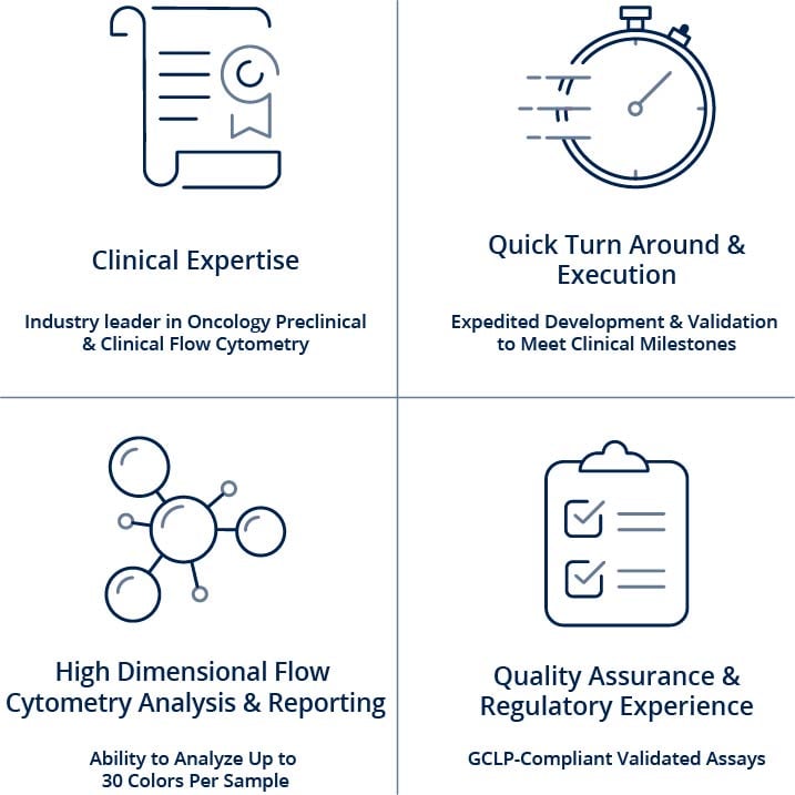
Champions' GCLP-compliant clinical flow cytometry services are implemented using fully optimized and validated human panels developed to meet your clinical trial needs.
We understand that every flow cytometry assay is unique, and therefore, we offer custom assay development and fit-for-purpose validation to suit our clients' needs. Champions is a leader in high dimensional flow cytometry and knows how important receiving high-quality, reproducible data is to you – we do our best to do it right, with scientific integrity.
Champions leads the industry in clinical flow cytometry expertise and innovative technology and will provide you with the highest quality, complex computational results with the ability to interrogate up to 30 parameters on each individual cell simultaneously within every patient sample. We also have experience executing challenging flow cytometry-based receptor occupancy (RO) assays, intracellular cytokine assays (ICS), and phosphorylation-based functional assays.


In vivo models for numerous diseases and conditions have endpoints that have involved animals being falling ill or dying. As researchers, we have sought to use animal models in more humane and practical ways using surrogate endpoints that have been developed to prevent animals from suffering while still providing critical research data. Flow cytometry has been instrumental in these advances.

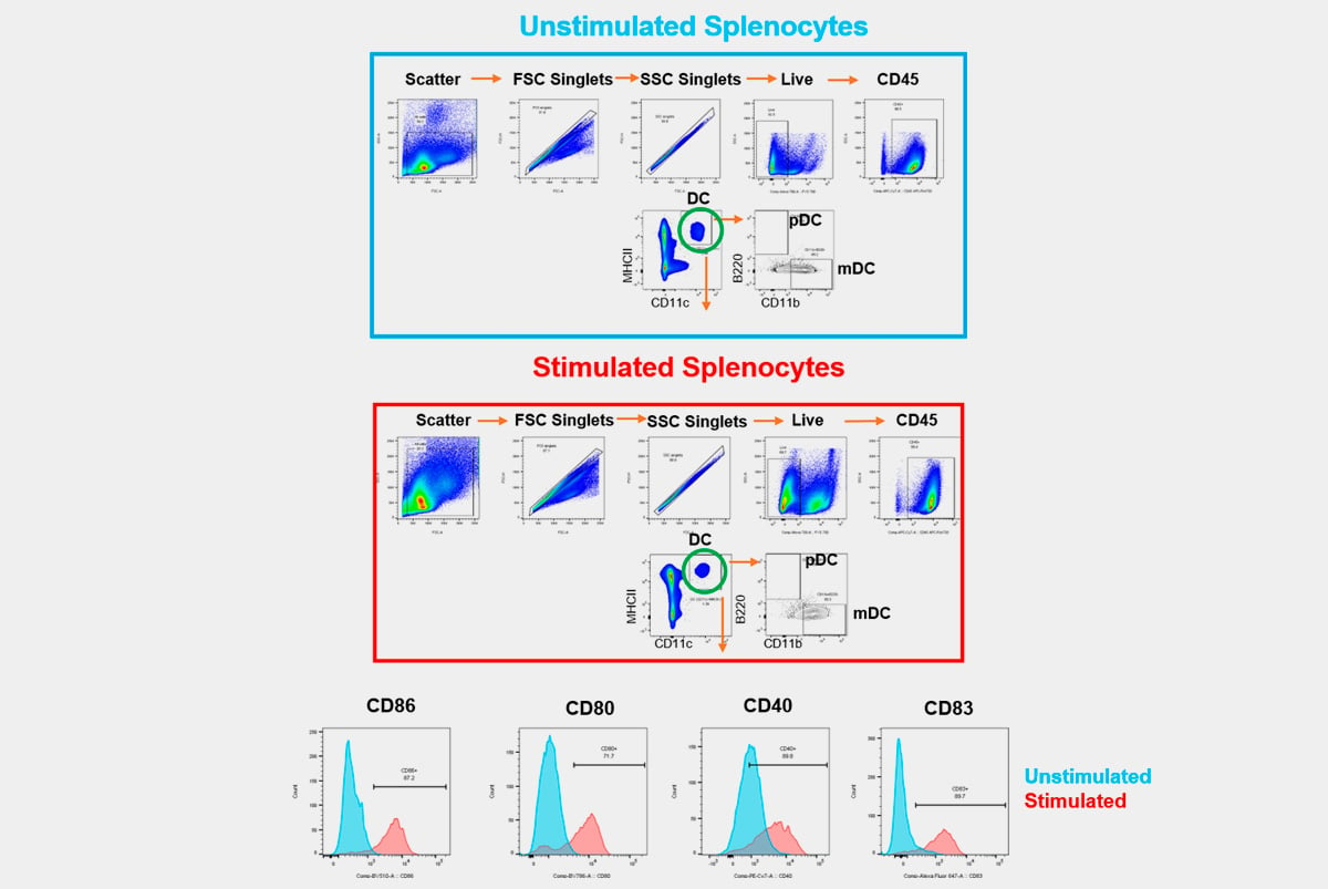
Immunotherapies are focused on strategies that alter immune responses, using antibodies that bind to receptors on different immune cell subsets to activate or suppress immune cell function. As such, it was necessary to develop assays that assess the functional and biological effect of a therapeutic on its target. When incorporated into high-parameter flow cytometry panels, receptor occupancy assay can simultaneously evaluate receptor expression and drug occupancy on defined cell subsets, to provide functional effects and safety information.