DNA Damage Assays
Automated DNA damage assays to study cellular DNA damage and repair mechanisms induced by cancer-causing genotoxic mutagens.
- Analytical Assays For Endpoint Data Collection
- DNA Damage Assays

Choose the Best DNA Damage Assays for your Preclinical Study
Champions offers three different methods for studying DNA damage and repair in tumor cells, each with different applications and advantages.
-
Comet assay to detect DNA single/double strand breaks in single cells
-
γH2AX IHC staining to detect and localize DNA breaks
-
γH2AX detection by Western Blot to measure DNA breaks
Advanced Comet Assay for thorough assessment of DNA damage
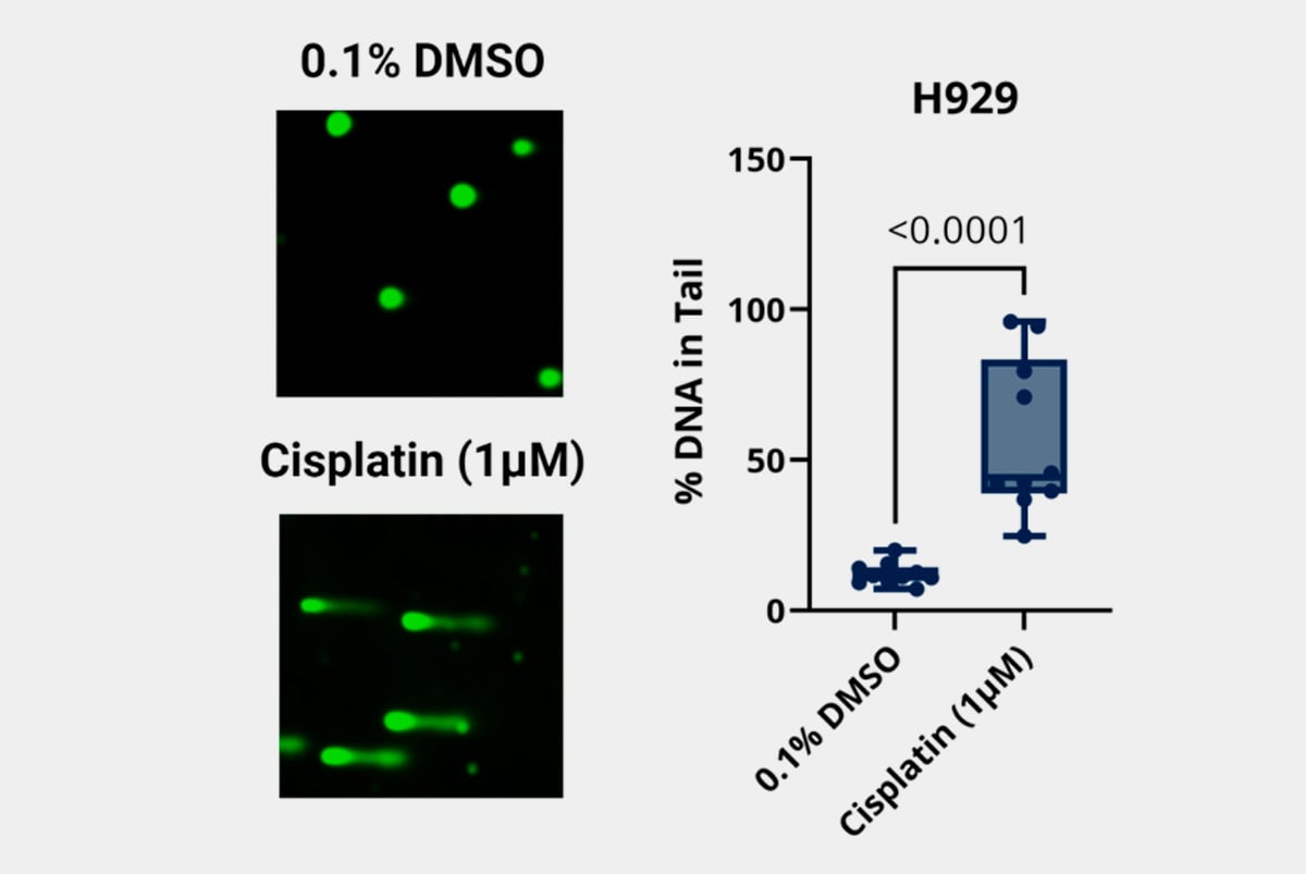
The Comet Assay is a sensitive and high-throughput technique for detecting DNA damage and repair at the single-cell level. Based on the Single-Cell Gel Electrophoresis (SCGE) principle, it identifies single-strand breaks (SSBs), double-strand breaks (DSBs), alkali-labile sites, and oxidative DNA damage. The assay exploits the ability of negatively charged DNA fragments to migrate through an agarose gel under an electric field. Cells are treated with a lysis solution to remove membranes, cytoplasm and nucleoplasm, leaving behind a nucleoid mass. During electrophoresis, damaged low-molecular-weight DNA fragments move toward the anode, forming a "comet tail"—with longer tails indicating greater DNA damage.
-
Comet assay to detect DNA single/double strand breaks in single cells
-
Measure DNA strand breaks with high sensitivity
-
Measure activity of agents involved in DNA damage repair
γH2AX Staining Assay to quantify of DNA damage & genomic instability
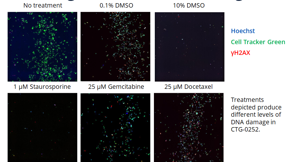
The γH2AX Staining Assay is a widely used method for detecting DNA double-strand breaks (DSBs), a key indicator of genomic instability. When cells are exposed to DNA-damaging agents, H2AX histone proteins become phosphorylated, forming γH2AX foci at break sites. These foci can be detected using immunostaining techniques or Western blot analysis to assess DNA damage. This assay is essential in genotoxicity testing (evaluating DNA damage from drugs, radiation, and chemicals), cancer research (analyzing DNA repair mechanisms and treatment responses), and as a biomarker for DNA damage response (studying genomic instability and aging).
-
Quantify DNA double strands breaks and damage
-
Genotoxicity testing (measuring DNA damage and repair from drugs, radiation, and chemicals)
-
Biomarker for DNA damage response measuring Measure genomic instability and telomere dysfunction
What are DNA damage assays used for?
DNA damage assays are laboratory techniques used to detect and quantify damage to DNA caused by environmental factors, radiation, chemicals, or biological agents. These assays are essential in toxicology, cancer research, genotoxicity testing, and drug development.
Key Applications:
- Genotoxicity Testing – Evaluating the effects of chemicals, drugs, radiation, and environmental toxins on DNA integrity.
- Cancer Research – Studying DNA repair mechanisms, genomic instability, and the impact of anti-cancer therapies.
- Drug Development & Toxicology – Assessing the safety and potential mutagenicity of new pharmaceuticals.
- Radiation Biology – Measuring DNA damage and repair following exposure to ionizing radiation.
- Biomarker Development – Using DNA damage markers (e.g., γH2AX) to assess disease progression and treatment response.
These assays help in understanding genetic stability, disease mechanisms, and the impact of external stressors on DNA, making them crucial in cancer research.
What methods are commonly employed to detect DNA damage?
Several techniques are used to detect and quantify DNA damage, each with specific applications in research, toxicology, and medical diagnostics. Below are some of the most commonly employed methods:
1. Comet Assay (Single-Cell Gel Electrophoresis, SCGE)
- Detects single-strand breaks (SSBs), double-strand breaks (DSBs), and oxidative DNA damage.
- Uses electrophoresis to separate fragmented DNA, forming a "comet tail" in damaged cells.
2. γH2AX Staining Assay
- Detects double-strand breaks (DSBs) by identifying phosphorylated H2AX histone (γH2AX foci).
- Visualized using immunofluorescence microscopy, flow cytometry, or Western blotting.
3. TUNEL Assay (Terminal deoxynucleotidyl Transferase dUTP Nick End Labeling)
- Identifies DNA fragmentation, mainly used to study apoptosis (programmed cell death).
- Uses fluorescently labeled nucleotides to tag broken DNA ends.
4. Micronucleus Assay
- Detects chromosomal damage, breaks, and loss by identifying small, extra nuclei (micronuclei) in cells.
- Commonly used for genotoxicity and mutagenicity testing.
5. Ames Test
- Detects mutagenic potential of chemicals by measuring DNA mutations in bacteria.
- A widely used test for genotoxicity screening in drug development.
6. Chromosomal Aberration Assay
- Identifies structural chromosomal changes, including deletions, translocations, and duplications.
- Conducted in metaphase cells under a microscope.
7. 8-Oxo-dG Assay
- Measures oxidative DNA damage by detecting 8-oxo-7,8-dihydro-2′-deoxyguanosine (8-oxo-dG), a marker of oxidative stress.
8. Alkaline and Neutral Gel Electrophoresis
- Alkaline Electrophoresis – Detects single-strand breaks and alkali-labile sites.
- Neutral Electrophoresis – Identifies double-strand breaks.
These methods provide insights into DNA integrity, damage response, and repair mechanisms, playing a crucial role in genotoxicity testing, cancer research, and drug safety assessment.
How does the comet assay detect DNA damage?
The Comet Assay (Single-Cell Gel Electrophoresis, SCGE) detects DNA damage at the single-cell level by measuring DNA strand breaks and fragmentation. It works by exploiting the electrophoretic mobility of negatively charged DNA fragments, which migrate through an agarose gel in response to an electric field.
Detection Process:
- Cell Embedding – Cells are mixed with agarose and spread on a microscope slide.
- Cell Lysis – The slide is treated with a lysis solution to remove cell membranes and proteins, leaving only the DNA structure (nucleoid).
- Electrophoresis – Under an alkaline or neutral buffer, an electric field is applied:
- Damaged DNA (strand breaks) has small, fragmented pieces that migrate toward the anode, forming a "comet tail."
- Intact DNA remains in the head region of the comet.
- Staining & Imaging – DNA is stained with a fluorescent dye (e.g., ethidium bromide or SYBR Green) and visualized under a fluorescence microscope.
- Analysis – The length and intensity of the comet tail correlate with the extent of DNA damage—longer tails indicate more damage.
What is the significance of γ-H2AX in DNA damage assessment?
γ-H2AX is a gold-standard marker for detecting and quantifying DNA double-strand breaks, making it an essential tool in genotoxicity studies, cancer research, and drug development.
γ-H2AX (phosphorylated H2AX at serine 139) is a key biomarker for DNA double-strand breaks (DSBs), one of the most severe types of DNA damage. When DSBs occur, the H2AX histone protein is rapidly phosphorylated, forming γ-H2AX foci at the damage sites. These foci can be detected using immunostaining, flow cytometry, or Western blotting, making γ-H2AX a highly sensitive marker for DNA damage assessment.
Importance in DNA Damage Studies:
- Early and Sensitive DNA Damage Indicator – γ-H2AX is one of the first responses to DSBs, appearing within minutes of damage.
- Quantification of DNA Damage – The number and intensity of γ-H2AX foci correlate with DSB severity and cellular stress levels.
- Genotoxicity Testing – Used to evaluate the DNA-damaging effects of radiation, chemicals, and drugs.
- Cancer Research – Helps study DNA repair mechanisms, tumor progression, and response to therapies like radiation and chemotherapy.